| |
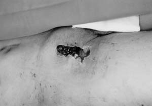 |
| Fig. 1 |
The area of skin pigmentation is swollen and pulsating. The
skin defect in the right groin area is the hemorrhagic lesion. |
|
| |
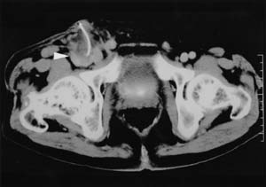 |
| Fig. 2 |
Enhanced computed tomography shows the right femoral pseudoaneurysm.
Culture of perianeurysmal tissue, seen has as a vague mass (arrow), was positive
for methicillin resistant S. aureus. |
|
| |
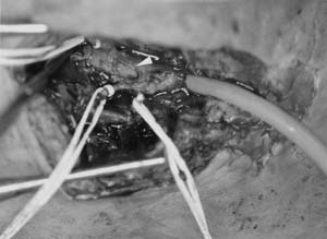 |
| Fig. 3 |
The surgical view shows a ruptured pseudoaneurysm (arrow) of
the right common femoral artery. |
|
| |
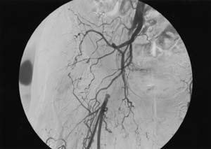 |
| Fig. 4 |
Postoperative arteriogram shows good collateral blood flow between
the right common iliac artery and profound and superficial femoral arteries. |
|
|