| |
 |
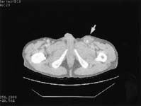
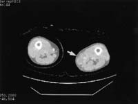
| Fig. 1 |
Preoperative CT findings. a: groin slice. b: above-knee popliteal
fossa slice. Arrow indicates the abscess formation around the prosthesis. |
|
| |
|
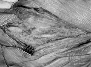
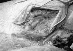
| Fig. 2 |
Intraoperative photographs of the groin. a: Proximal part of
sartorius muscle flap is dissected. b: the graft is wrapped with the muscle flap. |
|
| |
|
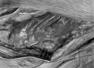
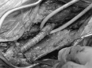
| Fig. 3 |
Intraoperative photographs of the above-knee popliteal fossa.
a: Distal part of sartorius muscle flap is dissected. b: the graft is wrapped
with the muscle flap. |
|
|