| |
|
|
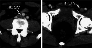
| Fig. 1 Preoperative enhanced computed tomography. lt.
CIV: left common iliac vein, lt.CFV: left common femoral vein |
|
| |
| |
|
|
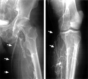
| Fig. 2 Preopeative venography. →: left great saphenous
vein |
|
| |
| |
|
|
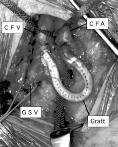
| Fig. 3 Operative photograph of the A-V fistula. CFA:
common femoral artery, CFV: common femoral vein, GSV: great saphenous vein |
|
| |
| |
|
|
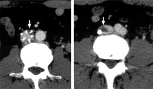
| Fig. 4 Postoperative enhanced computed tomography. →:
intravenous thrombus entrapped by temporary IVC filter |
|
| |
| |
|
|
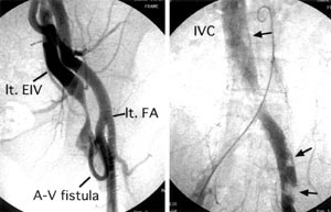
| Fig. 5 Postoperative angiography. →: thrombus,
lt. EIV: left external iliac vein, lt.FA: left femoral artery. |
|
| |
|