| |
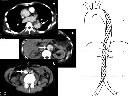 |
| Fig.
1 |
Enhanced
computed tomography (CT) at re-dissection showed new
blood lumen (white arrows) and the aorta was partially
three chambered (slice A). The superior mesenteric artery
was enhanced and the true lumen was compressed with
the pseudolumen (slice B). Each CT image was orientated
as right side schematic drawing alphabetically (the
oblique lined area shows pseudolumen). |
|
| |
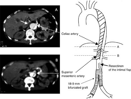 |
| Fig.
2 |
Post
operative enhanced computed tomography (CT) showed blood
flow to the celiac artery from true lumen and to the
superior mesenteric artery from the pseudolumen (white
arrows). Each CT image was orientated as right side
schematic drawing alphabetically (the oblique lined
area shows pseudolumen). |
|
|